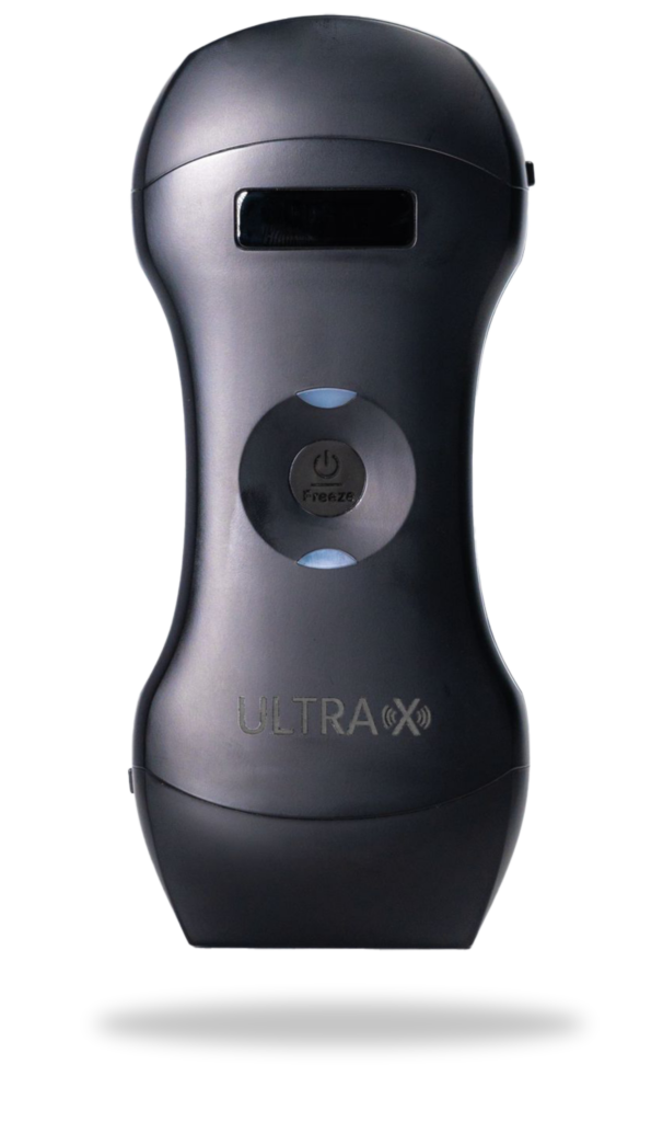
Probe hand-piece
Dual-head wireless handheld ultrasound scanner

Dual-head wireless handheld ultrasound scanner
Advanced digital imaging technology, clear image
Wireless connectivity, easy to operate
Built-in and replaceable battery
Cost-effective, small and light, easy to carry
Applicable in hospital, emergency, clinic, outdoor


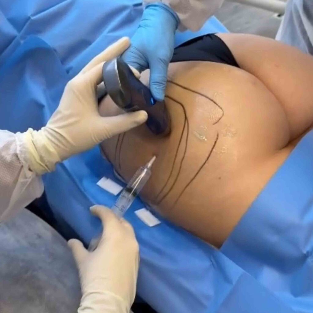
ULTRA X Ultrasound Dual Doppler technology plays a crucial role in aesthetic practices by providing detailed insights into vascular structures.
Practitioners often use it for vein mapping before cosmetic procedures, ensuring precise identification of veins to avoid inadvertent injections.
Additionally, Ultra X Doppler ultrasound assists in assessing blood flow, particularly valuable in treatments involving vascular structures like facial rejuvenation or sclerotherapy.
This technology enhances procedural safety, offering a visual guide to practitioners, helping them navigate and understand vascular anatomy, and ultimately minimizing the risk of complications during aesthetic interventions.

Probe Frequency:
Convex head: 3.5MHz/5MHz
Linear head: 7.5MHz/30MHz
Probe Element: 128
Scan Depth:
Convex head: 90/160/220/305mm
Linear head: 20/40/60/100mm
Head Width:
Convex head: 45mm
Linear head: 40mm
Harmonic, Denois
Weight: 260g (0.6 lbs)
Dimension: 156mm x 60mm x 20mm
(6.1″ x 2.4″ x 0.8″)
Battery Life: 22.5 hours
Charger: wireless
Recharge Time: fully charged within 2 hours
Working system: Apple iOS and Android
Scanning mode: Electronic array
Display mode: B,B/M;
Colour Dopple: B+colour, B + PDI,B+PW
Measure: Length, Area, Angle, Heart Rate,
Obstetrics Format: jpg, avi, and DICOM
Frame Rate: 18 frames/second
Image Adjust: BGain, TGC, DYN, Focus,Depth,